Skip to content
DN2M
Annuaire
Navigation
anpaa-bretagne.fr
sportmag.info
blog-sport.net
belle-et-epanouie.fr
touslesfaits.fr
lejardindegaia.fr
medialibre.fr
mariage-univers.fr
lespecialistedumariage.fr
passion-animaux.net
vayavoirdusport.fr
leblogmariage.fr
nouvelle-dimension.fr
immowebpartner.fr
mistercash.net
info-immobilier.net
lescoudes-surlatable.fr
wedding-news.net
blog-mariage.net
animal-passion.net
labonnemaison.fr
beauty-inc.net
notresweethome.com
bertrandbarre.com
paranormalnews.fr
mordudesport.fr
mariage-magazine.fr
univers-beaute.net
mon-animal-de-compagnie.net
actu-animaux.fr
actu-buzz.net
123mariages.com
monde-gourmandises.net
mamaison.info
partenaire-financier.com
actu-auto-buzz.fr
belle-et-bien.fr
blogueur.net
h-immobilier.fr
conseils-mariage.net
belle-et-naturelle.fr
animaleo.net
animal-news.fr
fashion-blog.fr
lentracte-gourmand.fr
dame-jeanne.fr
domaine-hebergement-ecommerce-serveur.fr
be-at-home.fr
univers-mariage.net
touslesanimaux.net
lemondedumariage.fr
easy-cooking.fr
revuerepublicaine.fr
sos-technologies.net
univers-animaux.net
espace-mariage.fr
planete-beaute.com
partimmobilier.fr
armoricauto.com
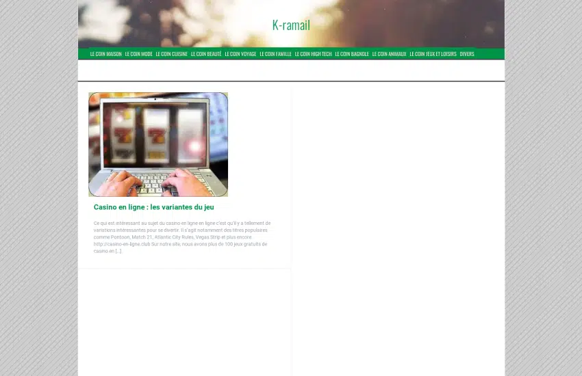
k-ramail.net
esprit-sport.com
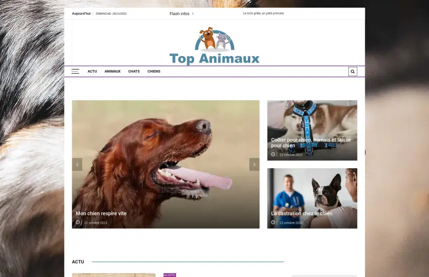
top-animaux.info
espace-beaute.net
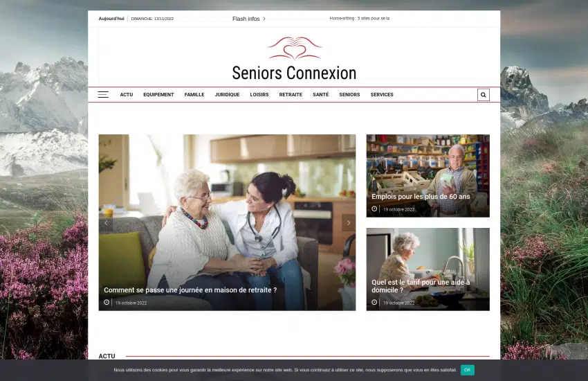
seniorsconnexion.fr
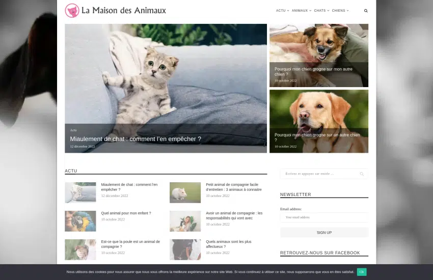
lamaisondesanimaux.net
leschiensnefontpasdeschats.fr
unefamille.net
coeurdemariage.fr
partir-en-classe.org
diversite-et-emploi.fr
cuisine-gratuite.com
acticarriere.fr
no-passion.com
info-mariage.net
vismaviedesenior.fr
lemondedesanimaux.info
lemondedusport.fr
conseils-beaute.com
la-une-des-journaux.info
planifiez-votre-mariage.fr
yadusport.com
lebloginfo.fr
la-maison.info
noxautos.fr
avantage-seniors.fr
format-sport.com
au-comptoir-immobilier.com
atmospheredujardin.com
espace-senior.info
jeune-senior.net
seniors-univers.fr
cercle-des-seniors.fr
mariage-conseils.fr
beaute-unique.fr
sport-univers.fr
top-beaute.com
amazingpetplace.com
leblogdevoyage.fr
animal-liberation.net
lartdugout.fr
yakar.fr